


Osteoarthritis: what is it in simple words?
Osteoarthritis is a chronic pathology in which the gradual destruction of the cartilaginous plate occurs. Pathological changes affect the underlying bone, which becomes more compact and marginal growths (osteophytes) develop. The joint capsule reacts to events that occur and a reactive vasculitis develops.
Information about the disease and possible complications
The incidence of the pathology depends on age. The first signs of arthrosis usually appear no earlier than 30-35 years of age, and by the age of 70 about 90% of the population suffers from this pathology. Osteoarthritis has no gender differences. The only exception is degenerative damage to the joints between the carpal phalanges. This form of the disease is 10 times more common in women than men. Osteoarthritis most often affects the large joints of the legs and arms.
The pathological process begins with the interstitial substance of cartilage tissue, which includes type 2 collagen fibers and proteoglycan molecules. The normal structure of the interstitial substance is maintained by balancing the processes of anabolism and catabolism. If the process of breakdown of cartilaginous tissue dominates its synthesis, conditions are created for the development of osteoarthritis. This explains in simple terms what osteoarthritis is.
Most often, the first signs of the disease develop in places of greatest mechanical load, with the appearance of limited areas of softening of the cartilaginous plate. As the pathological process progresses, the cartilage fragments and cracks, and local deposition of calcium salts is possible. Beneath the cartilaginous defects the underlying bone is exposed; Separate fragments of cartilage enter the joint cavity and can lead to the so-called "jam" (symptoms of a "joint mouse").
Damage to the cartilage lining the articular processes of the bones leads to the fact that they lose their ideal shape, repeating the contours of each other. As a result, during displacement, the joint surfaces experience a non-physiological load. In response to this, compensatory resynthesis processes are stimulated in the bone tissue. The bone becomes denser (subchondral osteosclerosis develops) and irregularly shaped marginal growths (osteophytes) appear, which further changes the discrepancy between the articular surfaces. The development of pathological changes gradually limits the range of motion of the joint and contributes to the development of complications in the form of muscle contractures (secondary muscle spasm that occurs in response to pain).
Osteoarthritis becomes the background for the development of synovitis - inflammation of the synovial membrane of the joint. This is due to the fact that dead fragments of cartilage and bone activate phagocytic leukocytosis, which is accompanied by the release of proinflammatory mediators. Over time, such long-term inflammation is accompanied by sclerosis of the periarticular tissues: the joint capsule thickens, the surrounding muscles atrophy.
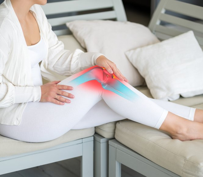
The main symptom of osteoarthritis is pain, which over time is accompanied by limited mobility of the joint. The limitation of mobility is first of a functional compensatory nature, then due to organic changes. Additional diagnostic imaging methods (X-ray, ultrasound, computed tomography or magnetic resonance imaging) help establish the correct diagnosis.
Depending on the stage and degree of arthrosis, treatment can be carried out with conservative or surgical methods. An orthopedic traumatologist will help you choose the optimal treatment program that takes into account the individual characteristics of the patient.
Types of osteoarthritis
There are 2 types of osteoarthritis:
- The primary variant is a consequence of a violation of the relationship between the processes of synthesis and degeneration in cartilage tissue and is accompanied by a disorder of the function of chondrocytes, the main cells of cartilage.
- The secondary variant occurs in a previously changed joint when the normal relationship (congruence) of the joint surfaces is disturbed, followed by a redistribution of the load on them and with a concentration of pressure in certain areas.
Symptoms of joint arthrosis
The main symptom of joint arthrosis is pain. It has some distinctive features that allow the primary diagnosis of the disease.
- Mechanical pain, caused by the loss of the shock-absorbing characteristics of the cartilage. Painful sensations occur during physical activity and are relieved during rest.
- Night pain.Caused by the stagnation of venous blood and the increase in blood pressure flowing inside the bone.
- Initial pain.It is short-lived and appears in the morning when a person gets out of bed (the patient says that he needs to "disperse"). These pains are caused by the deposition of debris on the cartilaginous plates; during the movement, these fragments are pushed into the joint inversions, so the unpleasant sensations cease.
- Meteor addiction.The pain may intensify when weather conditions change (increased atmospheric pressure, cold weather, excessive humidity).
- Blockage pain.These are sudden painful sensations associated with pinching of a fragment of bone or cartilage between the joint surfaces. Against the background of "blockage", the slightest movements in the joint stop.
The nature of the pain changes slightly when secondary synovitis occurs. In this case, the pain becomes constant. In the morning, a person is bothered by joint stiffness. Signs of the inflammatory process are determined objectively: swelling and local increase in skin temperature.
Osteoarthritis usually begins slowly with the onset of pain in an affected joint. At first the pain only bothers you during physical activity, but then it also appears at rest and during night sleep. Over time, pain is also felt in the joints on the opposite side, associated with a compensatory increase in load. An important distinguishing feature of arthrosis is its frequency, when short periods of exacerbation are followed by periods of remission. The progression of the pathological process is indicated by a reduction in the period of relapse and the development of adverse consequences in the form of contractures and a strong limitation of the mobility of the joint.
Course of arthrosis during pregnancy
During pregnancy, osteoarthritis can manifest itself in different ways. Usually, up to 12-13 weeks, an exacerbation of the pathological process associated with hormonal changes occurring in the woman's body may occur. The second and third trimesters are generally relatively stable. Management of pregnancy is carried out by an obstetrician-gynecologist and an orthopedic traumatologist.
Causes of joint arthrosis
The main mechanism that triggers the destruction of cartilage is a violation of the synthesis of proteoglycan molecules by cartilage tissue cells. The development of arthrosis is preceded by a period of metabolic disorders, which occurs in a hidden manner. This metabolic imbalance is characterized by damage to proteoglycans and their components (chondroitin, glucosamine, keratan), accompanied by disintegration and breakdown of the cartilage matrix. Collagen fibers break down in the cartilage plate, the supply of metabolites necessary for life is disrupted, and the water balance also changes (first the cartilage is hydrated, then the number of water molecules decreases sharply, which further stimulates the break).
Primary pathological processes negatively affect chondrocytes, which are very sensitive to the surrounding matrix. Changes in the qualitative characteristics of chondrocytes lead to the synthesis of defective proteoglycan molecules and short chains of collagen fibers. These defective molecules do not bind well to hyaluronic acid, so they quickly leave the matrix. With osteoarthritis, a "boom" of cytokines is also observed: the released cytokines interrupt the synthesis of collagen and proteoglycans and also stimulate inflammation of the synovial membrane.
The main causes of arthrosis can be various:
- "excess" weight, which increases the load on the joints;
- wearing low-quality shoes;
- concomitant diseases of the musculoskeletal system;
- suffered joint injuries.
Signs and diagnosis of joint arthrosis
Based on the clinical symptoms, the radiologist makes a preliminary diagnosis. To confirm this, further imaging tests are performed.
- X-ray.At an early stage, radiographic signs of the disease are of little importance: they may be an uneven narrowing of the joint space, slight compaction of the underlying bone and small cysts in this area. At a later stage, the x-ray is more informative: marginal bone growths appear, the shape of the joint surfaces changes, joint "mices" and areas of calcification in the capsule can be determined.
- Ultrasound of the joints.Ultrasound scanning is more informative for detecting early signs of osteoarthritis. Signs such as intra-articular effusion, changes in the thickness and structure of the cartilaginous plate, and secondary reactions of the capsule, musculotendinous, and ligament compartments may be seen.
- Computed or nuclear magnetic tomography.This diagnosis of joint arthrosis is carried out in complex clinical cases, when it is necessary to evaluate in detail the condition of the cartilaginous plate, the subchondral region of the bone and determine the volume of synovial fluid, incl. in joint inversions.
Expert opinion
Deforming arthrosis of the joints is one of the most common pathologies of the musculoskeletal system, affecting 10-15% of the world's population. The insidiousness of the disease is that it develops slowly and gradually. Initially, these are short-term pains in a joint, which a person often does not pay attention to. Gradually, the severity of the pain syndrome becomes more intense, while the periodic nature of the pain turns into constant. In the absence of treatment, the disease continues to progress and is accompanied by severe degeneration of the cartilage, which no longer responds to conservative therapy and to solve this problem only arthroplasty is necessary, a complex and expensive operation to replace the destroyed joint with a complete denture. full-fledged system. However, targeted drug therapy and lifestyle modification can help significantly delay this or avoid it altogether. Therefore, if you experience joint pain, it is important to visit a doctor as soon as possible.
Treatment of osteoarthritis
According to clinical guidelines, the main goal of osteoarthritis treatment is to slow the progression of degenerative lesions of the cartilaginous plate. To achieve this, measures are taken that reduce the load on the damaged joint and promote its recovery, and therapy is prescribed to stop the development of secondary synovitis.
Conservative treatment
Unloading the joint is obtained in the following ways:
- loss of body weight (if it is excess);
- perform physical therapy that excludes prolonged similar poses;
- refusal to lift large loads or remain kneeling for long periods of time (relevant for some professions).
In the initial stages of the disease, in addition to physiotherapy, swimming and cycling are useful. In the later stages, to unload the joint during an exacerbation, it is recommended to walk with an orthopedic stick or use crutches.
For pain relief, incl. against the background of secondary synovitis, nonsteroidal anti-inflammatory drugs are used, both local and systemic. Intra-articular injections of corticosteroids can be used for the same purpose.
To improve the anatomical and functional state of the cartilaginous plate, chondroprotectors and hyaluronic acid preparations are used, which are injected into the joint cavity. They help improve the metabolism of cartilage tissue, increase the resistance of chondrocytes to damage, stimulate anabolic processes and block catabolic reactions. This allows you to slow down the progression of the pathological process and improve the mobility of the joint.
Surgery
Surgical treatment options depend on the stage and activity of the disease process.
- Joint puncture– indicated for severe reactive synovitis. It allows not only to remove the inflammatory fluid, but also to introduce corticosteroids that interrupt the pathological chain.
- Arthroscopic interventions, which involve the introduction of instruments into the joint cavity through small punctures and subsequent visualization under magnification. These interventions allow you to wash the joint and its inversions, level the cartilaginous plate, remove necrotic areas, "polish" the joint surfaces, etc.
- Endoprosthesis– is considered a radical operation, which is performed in case of an advanced pathological process. Typically used for osteoarthritis of the knee or hip joint.
Prevention of osteoarthritis
Prevention of arthrosis is aimed at maintaining a normal weight, wearing orthopedic shoes, avoiding work on the knees, dosed lifting of heavy objects and compliance with a physical activity regime.
Rehabilitation for joint arthrosis
Rehabilitation for joint arthrosis involves a set of procedures capable of improving the functional state of the joint and surrounding tissues. Physiotherapy, therapeutic massage and healing gymnastics are used.
Questions and answers
Which doctor treats osteoarthritis?
Diagnosis and treatment are carried out by a traumatologist-orthopedist.
Does x-ray always allow the correct diagnosis to be made?
The severity of clinical signs of osteoarthritis does not always correlate with radiological changes. Often in practice there are cases when, with severe pain, the x-ray does not reveal significant changes, and, vice versa, when a "bad" x-ray image is not accompanied by significant symptoms.
Is diagnostic arthroscopy performed for osteoarthritis?
If arthrosis is suspected, arthroscopy is usually performed not to establish a diagnosis, but to look for causes that can lead to disruption of the functional state of the joint (for example, damage to the menisci of the knee joint and ligaments intraarticular).