


What is spinal osteochondrosis in simple words?
Spinal osteochondrosis is a chronic disease based on degenerative-dystrophic changes in the intervertebral disc resulting in involvement of adjacent vertebrae, intervertebral joints and spinal ligaments in the process.
The word “osteochondrosis” has two Greek roots: οστό - bone and χόνδρος - cartilage.
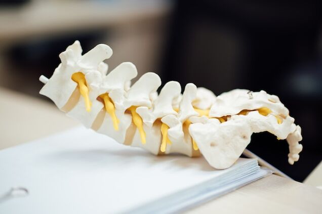
Vertebrae are formations made up of spongy bone.They are connected to each other by cartilaginous discs.There are ligaments along the anterior and posterior surfaces of the vertebrae.Cartilage discs prevent the vertebrae from coming together and the ligaments from moving apart.Thanks to the coordinated work of discs and ligaments, the spine is elastic, and this allows it to carry out vital functions:
- ensure balance in a vertical position,
- soften shocks and shocks when walking and jumping,
- protect the skull and the brain located in it from impacts due to excessive shocks.
With osteochondrosis, protrusions of the intervertebral discs form beyond the vertebral bodies.Depending on the direction in which the protrusion occurs and its size, pain, numbness, muscle discomfort and other symptoms develop.
ICD-10 codes:
- M42 Osteochondrosis of the spine
- M42.0 Juvenile osteochondrosis of the spine
- M42.1 Osteochondrosis of the spine in adults
- M42.9 Osteochondrosis of the spine, unspecified
- M43.1 Spondylolisthesis
- M47 Spondylosis
- M47.0 Anterior spinal or vertebral artery compression syndrome
- M47.1 Other spondyloses with myelopathy
- M47.2 Other spondyloses with radiculopathy
- M48.0 Spinal stenosis
- M50.0 Damage to the intervertebral disc of the cervical spine with myelopathy
- M50.1 Damage to the intervertebral disc of the cervical spine with radiculopathy
- M50.2 Displacement of the intervertebral disc of the cervical spine of other types
- M50.3 Other degeneration of the cervical intervertebral disc
- M51.0 Lesions of the intervertebral discs of the lumbar region and other parts with myelopathy
- M51.1 Lesions of the intervertebral discs of the lumbar region and other parts with radiculopathy
- M51.2 Other specified intervertebral disc displacement
- M51.3 Other specified intervertebral disc degeneration
- M53 Other back disorders, not elsewhere classified
Types of osteochondrosis
Depending on the part of the spine where the changes occur, there are several variants of the disease:
- cervical,
- chest,
- lumbar,
- sacral,
- mixed variants (cervicothoracic, lumbosacral).
Depending on the duration of the symptoms, the disease can be:
- acute (up to 3 weeks),
- subacute (3-12 weeks),
- chronic (more than 12 weeks).
According to the predominant neurological manifestation:
- with myelopathy (damage to the spinal cord),
- with radiculopathy (crushed and inflamed nerve roots).
Causes of osteochondrosis
To date, there is no exact data on the causes of osteochondrosis.
The role of genetic predisposition, mechanical damage and inflammation is recognized in the appearance of premature wear of intervertebral discs.
Intervertebral discs have no blood or lymph vessels of their own.The vessels of the vertebrae play a role in their nutrition and cleansing of harmful substances.With age and/or exposure to harmful influences, blood and lymph flow decreases, the discs receive less oxygen and nutrients, and harmful substances can accumulate inside them.All this leads to progressive wear.The degree and speed of disc wear increases when exposed to risk factors.
Risk factors:
- congenital anomalies of the vertebrae and spinal canal;
- flat feet;
- occupational risks (vibrations, heavy lifting, prolonged stay in an uncomfortable and constrained position, exposure to toxic substances);
- sedentary lifestyle;
- obesity;
- an unbalanced diet in protein, fat, vitamin and mineral content;
- insufficient consumption of clean water;
- smoking;
- environmental pollution.
Symptoms of spinal osteochondrosis
Listed by frequency of occurrence:
- pain;
- decreased range of motion;
- numbness, loss of sensation;
- decreased muscle strength;
- dysfunction of organs whose innervation is associated with the problematic part of the spine.
Clinically significant manifestations of spinal osteochondrosis are observed in 51 people per 1000 inhabitants.
The location of pain and other symptoms depends on the problematic part of the spine.
Cervical osteochondrosis:
- pain in the arms, shoulders, neck, aggravated by turning and tilting the head;
- heachache;
- decreased muscle strength in the arm;
- noise in the head, dizziness, flashing of "floaters", colored spots before the eyes in combination with burning, throbbing headache (vertebral artery syndrome).
Brain health depends on the condition of the cervical spine, since the arteries leading to the brain pass through the canal formed by the processes of the vertebrae.If, due to osteochondrosis, the lumen of the canal narrows, blood flow through the arteries is disrupted and the brain experiences a lack of oxygen and nutrients.
Thoracic osteochondrosis:
- pain in the chest, under the shoulder blade, in the heart area, aggravated by turning the body, coughing, sneezing;
- dysfunction of the gallbladder, stomach, esophagus.
Lumbar and/or sacral osteochondrosis:
- pain in the lower back, back and side of the thigh;
- numbness in the toes;
- increased frequency of urination (10-12 times a day, perhaps more), involuntary loss of urine during physical activity;
- sexual disorders.
Due to frequent pain, half of people suffering from osteochondrosis show signs of constant emotional stress.
Stages of development and course of osteochondrosis
The initial stage of osteochondrosis is manifested by a dull, aching pain in the back or lower back that occurs during prolonged standing, after walking or running;neck pain, aggravated by turning and tilting the head.
As the disease progresses, the intervertebral disc or discs may swell (herniate) and, as a result, compress the nerve root (radiculopathy).This leads to intense pain radiating to the arm or leg, muscle weakness, disorders of skin sensitivity, vascular tone and the function of organs receiving innervation from the problem part of the spine.In more severe cases, compression of the spinal cord may occur, leading to paresis or paralysis.
Osteochondrosis is a chronic disease.After adequate treatment, remission occurs, that is, the symptoms decrease or disappear completely.If a new protrusion of the intervertebral disc forms, an aggravation occurs and pain and other symptoms return.
Diagnostics
Examination by a neurologist.
Basic instrumental research methods:
- magnetic resonance imaging (MRI),
- computed tomography (CT).
Further:
- spondylography (thorough x-ray examination of the spine),
- electromyography (EMG),
- electroneuromyography (ENMG),
- bone densitometry (performed to detect osteopenia/osteoporosis).
Basic laboratory methods:
- general blood test,
- general urinalysis,
- biochemical blood tests (glucose, creatinine, urea, electrolytes, bilirubin, liver and pancreatic enzymes; glycated hemoglobin, C-reactive protein),
- coagulogram.
Further:concentration of calcium and phosphates in the blood.
Treatment of osteochondrosis
Conservative treatment
It is performed if the patient does not have acutely progressive neurological symptoms.
Goals:
- reduction or relief of pain,
- correction of muscle tone,
- reduction of inflammation and swelling,
- prevent the progress of dystrophic changes in the structures of the spine,
- correction of impaired functionality of internal organs,
- increase the patient's daily activity,
- teach the patient to cope with pain.
Conservative treatment of osteochondrosis includes:
- respect for a rational motor regime,
- use of drugs,
- physiotherapy,
- massage,
- Physical therapy (after pain relief and stabilization of condition),
- acupuncture,
- manual therapy.
Pharmacological treatment
The main groups of drugs that can relieve or relieve pain and stabilize the condition of a patient with osteochondrosis are listed.Only a doctor can select an adequate treatment regimen, taking into account the characteristics of the clinical picture of a particular patient.
Non-steroidal anti-inflammatory drugs(NSAIDs):
- for oral administration,
- for intramuscular injections,
- for intravenous administration,
- for insertion into the rectum (rectal suppositories),
- for external use (ointment, gel).
Muscle relaxants(drugs that reduce muscle spasticity).
Used for severe tension and painful muscle spasms.
Diuretics(to reduce local swelling).
Drugs that improve the condition of cartilage tissue(chondroprotectors):
- sodium chondroitin sulfate,
- a combination of sodium chondroitin sulfate and glucosamine.
B vitamins:
- thiamine (B1),
- pyridoxine (B6),
- cyanocobalamin (B12),
- combination B1+B6+B12.
In the acute period, with severe pain, bed rest for 1-2 days is possible, which helps to relax the muscles and reduce pressure inside the cartilaginous disc.We recommend wearing a stabilizing lumbar corset or Shants collar.
As the intensity of the pain decreases, the treatment is supplemented with special therapeutic exercises aimed at stretching the spine and relaxing the muscles, with the gradual inclusion of exercises to form a muscle corset.Therapeutic manual massage is indicated.
With adequate therapy the pain gradually decreases and can disappear completely.There is also a regression of neurological symptoms.The improvement of the condition is caused by the decrease in the size of the herniated disc and the associated inflammatory changes in the surrounding tissues.
Surgical treatment
Emergency neurosurgical intervention is indicated for pelvic disorders with numbness of the anogenital area and ascending paresis of the feet (cauda equina syndrome).
The need for surgery may also arise if conservative therapy is ineffective within 3-6 months.
Prevent back pain
Avoid excessive physical activity (lifting heavy objects, carrying a heavy bag in one hand, etc.).
Avoid prolonged static loads (sitting, remaining in an uncomfortable position).
If your work involves such stress, it is recommended to take 10-minute breaks every 45 minutes, during which you need to walk.
Avoid hypothermia.
Maintain an adequate level of physical activity through regular exercise, swimming and/or walking.
Sleep on a medium-firm mattress.
Nutrition for osteochondrosis
A balanced diet and an adequate intake of liquids guarantee a normal supply of blood and nourishment to the vertebrae and, consequently, to the cartilaginous discs.As a result, metabolism and energy are normalized and harmful products do not accumulate.
Basic principles:
Daily calorie content, calculated individually, taking into account height, age, gender.
For overweight or obese patients, calorie intake should be limited.
Alcohol consumption regime– drink pure water, mineral waters and herbal teas in a volume of at least 1 liter per day, ideally at a rate of 30 ml/kg of body weight.
Daily use:
- wholemeal products (buckwheat, millet, oats);
- sufficient amount of protein (taking into account age and kidney function): animal - lean beef, chicken, turkey, rabbit, chicken egg (4-5 pieces per week);vegetables - beans, lentils, peas;
- healthy fats containing mono- and polyunsaturated fatty acids (fish, seafood, unrefined vegetable oils, unroasted and unsalted nuts, seeds);
- vegetables (both fresh and cooked), lettuce, herbs and leafy vegetables;
- berries - blueberries, blackberries, raspberries, cherries.
Exclusion from the diet:
- white bread and baked goods made from top quality flour;
- sugar, industrial sweets - sweets, cakes, biscuits, gingerbread, waffles;
- industrial drinks with added sugar - carbonated water, packaged juices;
- processed meat products: sausages, sausages, canned food.